Wednesday, February 7, 2018

How many cells are present in the body in an adult person?

A simple question deserves a simple answer. How many cells are in your body?
Unfortunately, your cells can’t fill out census forms, so they can’t tell you themselves. And while it’s easy enough to look through a microscope and count off certain types of cells, this method isn’t practical either. Some types of cells are easy to spot, while others–such as tangled neurons–weave themselves up into obscurity. Even if you could count ten cells each second, it would take you tens of thousands of years to finish counting. Plus, there would be certain logistical problems you’d encounter along the way to counter all the cells in your body–for example, chopping your own body up into tiny patches for microscopic viewing.
For now, the best we can hope for is a study published recently in Annals of Human Biology, entitled, with admirable clarity, “An Estimation of the Number of Cells in the Human Body.”

The authors–a team of scientists from Italy, Greece, and Spain–admit that they’re hardly the first people to tackle this question. They looked back over scientific journals and books from the past couple centuries and found many estimates. But those estimates sprawled over a huge range, from 5 billion to 200 million trillion cells. And practically none of the scientists who offered those numbers provided an explanation for how they came up with them. Clearly, this is a subject ripe for research.
If scientists can’t count all the cells in a human body, how can they estimate it? The mean weight of a cell is 1 nanogram. For an adult man weighing 70 kilograms, simple arithmetic would lead us to conclude that that man has 70 trillion cells.
On the other hand, it’s also possible to do this calculation based on the volume of cells. The mean volume of a mammal cell is estimated to be 4 billionths of a cubic centimeter. (To get a sense of that size, check out The Scale of the Universe.) Based on an adult man’s typical volume, you might conclude that the human body contains 15 trillion cells.

So if you pick volume or weight, you get drastically different numbers. Making matters worse, our bodies are not packed with cells in a uniform way, like a jar full of jellybeans. Cells come in different sizes, and they grow in different densities. Look at a beaker of blood, for example, and you’ll find that the red blood cells are packed tight. If you used their density to estimate the cells in a human body, you’d come to a staggering 724 trillion cells. Skin cells, on the other hand, are so sparse that they’d give you a paltry estimate of 35 billion cells.
So the author of the new paper set out to estimate the number of cells in the body the hard way, breaking it down into organs and cell types. (They didn’t try counting up all the microbes that also call our body home, sticking only to human cells.) They’ve scoured the scientific literature for details on the volume and density of cells in gallbladders, knee joints, intestines, bone marrow, and many other tissues. They then came up with estimates for the total number of each kind of cell. They estimate, for example, that we have 50 billion fat cells and 2 billion heart muscle cells.
Adding up all their numbers, the scientists came up with…drumroll…37.2 trillion cells.
This is not a final number, but it’s a very good start. While it’s true that people may vary in size–and thus vary in their number of cells–adult humans don’t vary by orders of magnitude except in the movies. The scientists declare with great confidence that the common estimate of a trillion cells in the human body is wrong. But they see their estimate as an opportunity for a collaboration–perhaps through an online database assembled by many experts on many different body parts–to zero in on a better estimate.

Wednesday, January 17, 2018

                              Biological Classification:-

Biological classification of Plants and Animals was first proposed by Aristotle on the basis of simple morphological characters.Linnaeus later classified all living organisms into two kingdoms:- Plantae and Animalia. R.H.whittaker proposed an elaborate five kingdom classification:- Monera, Protista, Fungi, Plantae and Animalia. The main criteria of the five kingdom classification were 1) Cell structure, 2) Body organization, 3) mode of nutrition, 4) Reproduction and 5) Phylogenetic relationships.


In the five-kingdom classification, bacteria are included in kingdom Monera. Bacteria are cosmopolitan in distribution. bacteria may be autotrophic or heterotrophic in their mode of nutrition.

Kingdom, Protista includes all single-celled Eukaryotes such as:- Chrysophytes, Dinoflagellates, Euglenoids, Slime-moulds, and Protozoans. Protista have defined Nucleus and other membrane-bound organelles. They reproduce asexually and sexually. 
 
Members of kingdom Fungi show a great Diversity in structure and habitat. Most fungi are saprophytic in their mode of nutrition. They show asexual and sexual reproduction.
 Ex:- Phycomycetes, Ascomycetes, Basidiomycetes, and Deuteromycetes as the four classes under this kingdom.

The Plantae includes all eukaryotic Chlorophyll-containing organisms. Algae, Bryophytes, pteridophytes, Gymnosperms, and Angiosperms are included in this group 

The heterotrophic eukaryotic, multicellular organisms lacking a cell wall are included in the kingdom Animalia. The mode of nutrition of these organisms is Holozoic. The reproduce mostly by the sexual mode.

Some Acellular organisms like viruses and viroids as well as the lichens are not included in the five-kingdom system of classification

Monday, January 15, 2018

Respiratory System:-

The process of exchange of O2 from the atmosphere with Co2 produced by the cells is called Breathing, commonly known as Respiration.

The respiratory system consists of the many parts like a Nose, pharynx(throat), larynx (voice box), trachea( windpipe), Bronchi and Lungs.

The Respiratory system has the following function:- 

1) Help in gaseous exchange- intake of oxygen for delivery to body cells and elimination of carbon dioxide produced by the cells.
2)Helps to regulate blood pH.
3)Contains receptors of a sense of smellProduced vocal sounds(phonation), and excretes the small amount of water and heat.Filter inspired air, 

Friday, January 12, 2018

Digestive System:

"The Breakdown of large insoluble of food molecules into the Small soluble molecules to be absorbed into the body cells".
The digestive system includes the Gastrointestinal (GI) tract and the accessory digestive organs. this GI or Alimentary canal is connected from mouth to anus by a tube.Organ of GI tract includes the mouth,
esophagus tube
stomach
the small intestine
the large intestine
most of the pharynx.
The accessory digestive organs include:-
The Teeth
Tounge
Salivary Gland
Liver
Gallbladder and
Pancreas.

Functions of Digestive system:-

 1) Ingestion:- taking food into the mouth
2)Secretion:- Release of water, acid, buffers, enzymes into the lumen of the GI tract.
3)Mixing and Propulsion:- Churning and propulsion of food through the GI tract.
4)Digestion:- The Mechanical and chemical breakdown of food.
5)Absorption:-passage and assimilation of digested product from GI tract into the blood and lymph.
6)Defection:- The elimination of feces from the GI tract.

Digestive Activities in the GI tract

 In the mouth:- The mouth is formed by the cheeks, hard and soft palates, lips and tongue.
Function:-Secrete Saliva from Salivary glands that softens moistens, and dissolves food and cleanses mouth and teeth.Salivary amylase present in saliva splits starch into smaller fragments.
                :-secrete Lingual lipase from lingual glands.
In Pharynx:- Receives a bolus from the oral cavity and passes it into the Esophagus tube. 
In Esophagus:- Receives a bolus from the pharynx and moves it into the stomach. The requires relaxation of the upper esophagus sphincter and secretion of mucus.
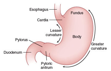
In Stomach:- The stomach has main four regions:-
1) The Cardia :- the superior opening area of the stomach are called Cardia.
2) Fundus:- To the left of the Cardia and rounder portion is called Fundus.
3) Body:- Inferior area and the large central portion is called Body. 
4) pylorus:- The region of the stomach that connects with the duodenum is The Pylorus.

Function of Stomach

1):-Mixes saliva, food and gastric juice to form chyme.
2):- Secretes Gastric juice, which contains HCl ( Secretion of Parietal cells that kills bacteria and denatures protein).
Pepsin( secretion of Cheif cells that begins the digestion of proteins)
Intrinsic factor( secretion of Parietal cells and aids absorption of vitamin B12) 
Gastric lipase( aids digestion of Triglycerides)
3):-Secretes gastrin( from G cells ) into blood which stimulates parietal cells to secrete HCL and Cheif cells to secrete Pepsinogen; contracts lower esophagus sphincter increases motility of the stomach and relaxes Pyloric Sphincter. 

The small intestine:-

Length 2-3 meter and in diameter 1-1.5 inches.
Regions of the small intestine include:-
1) The Duodenum:-C shape 
2) The Jejunum:- coiled shape
3) The Ileum:- Coiled shape
 

Function of the Small intestine:-

1)- Segmentation mix chyme with digestive juices and bring food into contact with the mucous for absorption; peristalsis propels chime through the small intestine.
2)- Completes the digestion of carbohydrates; proteins; and lipids; begins and completes and digestion of nucleic acids. 
3)-Absorbs about 90% of nutrients and water that pass through the digestive system.

The large intestine:-

The large intestine length 1-2 
Regions of the large intestine are the cecum, colon, rectum and anal canal.

Function of the large intestine:-

1) Haustral churning, peristalsis, and mass peristalsis drive the contents of the colon into the rectum.
2) Bacteria in the large intestine convert proteins to amino acids, breakdown amino acids and produce some B vitamins and vitamin k.
3)Absorbing some water, ions, and vitamins.
4) Forming feces.
5)defecating( emptying the rectum)


Thank you guys
#asthanigam97@gmail.com
if u have some doubt or any query then do a comment below and I'll solve it.
and please follow, comment and do #share...


Monday, January 8, 2018

#Enter-doudoroff pathway

Entner-Doudoroff Pathway

Entner-Doudoroff Pathway is an alternative pathway that catabolizes glucose to pyruvate using a set of enzymes different from those used in either glycolysis or the pentose phosphate pathway. This pathway, first reported by Michael doudoroff and Nathan Entner, occurs only in prokaryotes, mostly in gram-negative bacteria such as Pseudomonas aeruginosa, Azotobacter, Rhizobium.
                                In the pathway, glucose phosphate is oxidized to 2-keto-3-deoxy-6-phosphogluconic acid (KDPG) which is cleaved by 2-keto-3-deoxyglucose-phosphate aldolase to pyruvate and glyceraldehyde-3-phosphate. The latter is oxidized to pyruvate by glycolytic pathway where in two ATPs are produced by substrate-level phosphorylation. 

This process yields one ATP as well as one NADH and one NADPH for every glucose molecule.


Saturday, January 6, 2018

#CAM_Pathway

CAM pathway:-

Crassulacean acid metabolism(CAM) is a photosynthetic adaption in succulent (juicy and tasty) plants. Succulent plants, also known as Fat Plants, are xerophytic plants to arid climates or soil conditions. Succulent plants store water in their leaves and stems.The storage of water often gives succulent plants a swollen or fleshy appearance than other plants, a characteristic known as Succulence. The best-known Succulents are Cacti. these plants open their stomata during the night and close them stomata during the day. Closing stomata during the day helps succulent plants conserve water, and also prevents the Co2 from entering the leaves. During the night, when their stomata are open, these plants take up Co2. Assimilation of Co2 occurs into Malic acid at night which is stored in the vacuole. This mode of carbon fixation is called CAM (crassulacean acid metabolism). 

During the daytime, when the light rxn can supply ATP and NADPH for the Calvin cycle, Co2 is released from the malate for fixation through the Calvin cycle. This cycle differs from the C4 cycle. In C4 plants, the formation of the malic acids in the mesophyll is spatially separated from the Calvin cycle in the bundle sheath. In CAM plants, the two steps occur at separate times, but within the same cell.

Mechanism of CAM Pathway

In CAM pathway, formation of the C4 acids is temporally separated. This mech^ is following some steps which are given below:-
1)- At night, Co2 is captured by PEP carboxylase in the cytosol of mesophyll cell and PEP is carboxylated to oxaloacetate.
2)- The oxaloacetate is reduced to malate which is finally stored in the vacuole.Reduction of oxaloacetate is catalyzed by enzyme NAD+-malate dehydrogenase. 
3)- During the day, the stored malate transported back into the cytosol, where it is decarboxylated by an NADP+-malic enzyme. 
4)- The Co2 just released enters the chloroplast where it is fixed by the Calvin cycle.
"The adaptive advantage of CAM is the reduction of water loss by transpiration, achieved by the stomatal opening during the night".
#pathfinder 
#Asthanigam
#heplerblog
#tuneitfasthere
asthanigam97@gmail.com 
If u have any doubt or a query just comment below. I'll try solve it .
10q(thnku)


Friday, January 5, 2018


Chilling pictures from the early 1900s are the first crime scene photos ever… showing grisly murders as victims lie splayed in trashed rooms with blood ...

Love...Love

  " Every time I think of you, "A"       I feel the intensity of lovin' you      like you could never imagine.       ...